Full Paper: http://www.pnas.org/content/113/16/E2306.full
Researchers have turned fat cells into stem cells, which can then be programmed to repair any human tissue that was damaged by injury disease or ageing. The new breakthrough holds promising applications in treating neck pain, spinal injury, join and muscle degeneration and more.
| Live cell imaging of primary mature adipocytes harvested from Pdgfra-nGFP mice. The cells were treated with PDGF-AB (8 d)/AZA (2 d) and imaged over 8 d. GFP is expressed when Pdgfra–ve adipocytes transform into Pdgfra+ (MSC cell marker) cells. |
Abstract
Vashe Chandrakanthan et. al.
Current approaches in tissue engineering are geared toward generating tissue-specific stem cells. Given the complexity and heterogeneity of tissues, this approach has its limitations. An alternate approach is to induce terminally differentiated cells to dedifferentiate into multipotent proliferative cells with the capacity to regenerate all components of a damaged tissue, a phenomenon used by salamanders to regenerate limbs. 5-Azacytidine (AZA) is a nucleoside analog that is used to treat preleukemic and leukemic blood disorders. AZA is also known to induce cell plasticity. We hypothesized that AZA-induced cell plasticity occurs via a transient multipotent cell state and that concomitant exposure to a receptive growth factor might result in the expansion of a plastic and proliferative population of cells. To this end, we treated lineage-committed cells with AZA and screened a number of different growth factors with known activity in mesenchyme-derived tissues. Here, we report that transient treatment with AZA in combination with platelet-derived growth factor–AB converts primary somatic cells into tissue-regenerative multipotent stem (iMS) cells. iMS cells possess a distinct transcriptome, are immunosuppressive, and demonstrate long-term self-renewal, serial clonogenicity, and multigerm layer differentiation potential. Importantly, unlike mesenchymal stem cells, iMS cells contribute directly to in vivo tissue regeneration in a context-dependent manner and, unlike embryonic or pluripotent stem cells, do not form teratomas. Taken together, this vector-free method of generating iMS cells from primary terminally differentiated cells has significant scope for application in tissue regeneration.

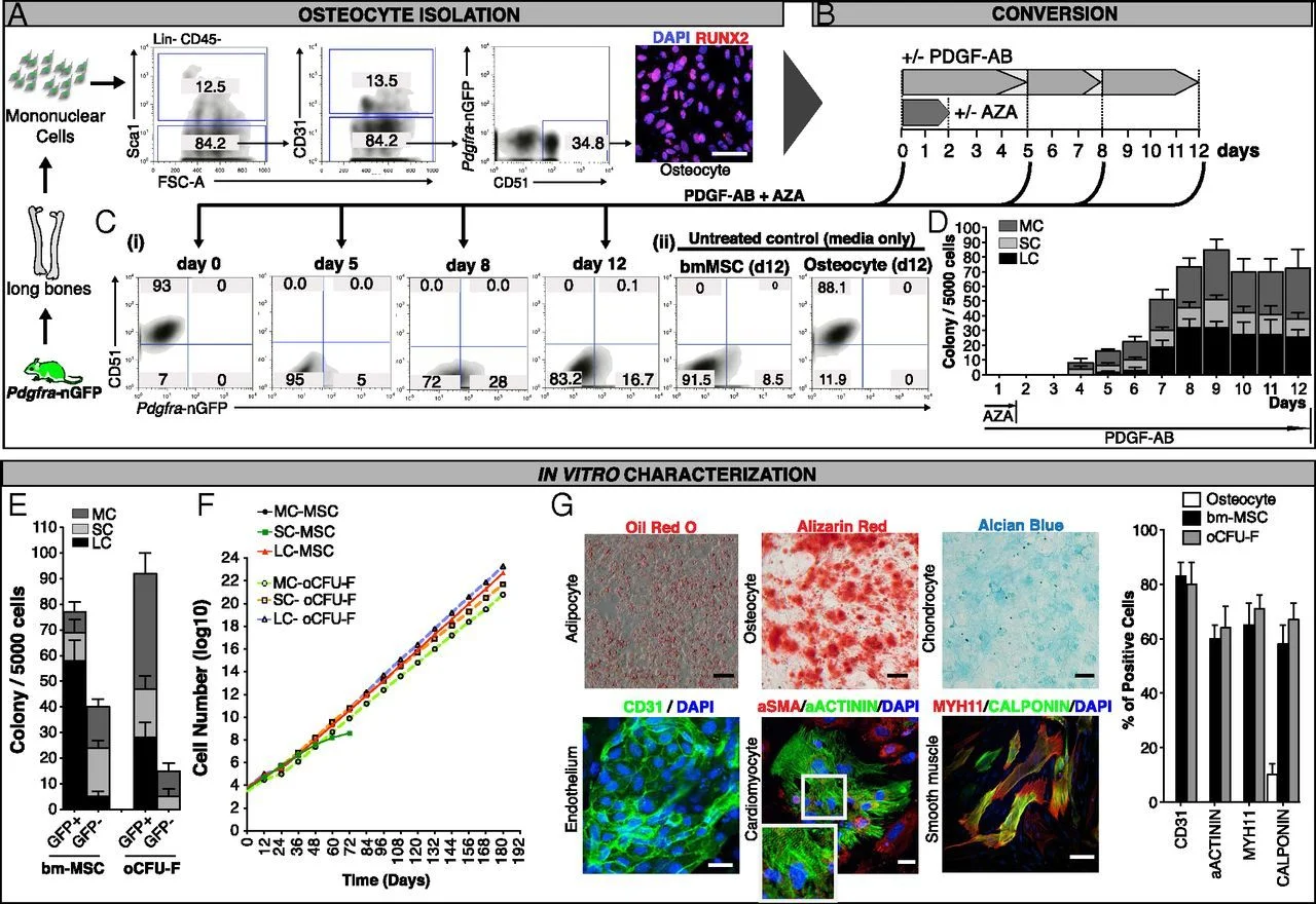
| Conversion of primary osteocytes from Pdgfrα-H2B:GFP mice into renewable multipotent cells. (A) Sorting strategy for harvesting Lin−/CD45−/SCA1−/CD31−/Pdgfrα-GFP−/CD51+ osteocytes from long bones of 8–16-wk-old mice. The Inset shows expression of RUNX2 by confocal immunofluorescence in sorted osteocytes. (B) Schematic outline of the treatment schedule. Osteocytes from A were treated and evaluated by flow cytometry (C) and CFU-F assays (D). (C, i) Flow cytometry profiles of freshly isolated Pdgfrα-GFP−/CD51+ osteocytes showing progressive loss of CD51 and acquisition of GFP when cultured in media containing PDGF-AB and AZA. (ii) Primary osteocytes treated with media alone did not show acquisition of GFP. A proportion of primary MSCs harvested from bone continue to express GFP. (D) Quantification of oCFU-F colonies (based on size) from osteocytes cultured in MSC medium supplemented for 1–12 and 1–2 d with PDGF-AB and AZA, respectively. (E) CFU-F colony number (based on size) derived from sorted bmMSCs and sorted oCFU-Fs. (F) Growth curves of micro-, small, and large colonies from bmMSCs and oCFU-Fs that were harvested at P1 and perpetuated in culture. (G) Histo- and immunohistochemistry showing multilineage differentiation potential of converted oCFU-Fs. The histogram to the Right shows a comparison of differentiation markers (see Lower panel of images) in untreated osteocytes, bmMSCs, and oCFU-Fs. Also see SI Appendix, Fig. S10 for an extended panel of tissue differentiation markers and quantification. bmMSCs, primary bone marrow mesenchymal stem cells; oCFU-F, colony-forming unit–fibroblasts derived from PDGF-AB/AZA-treated osteocytes. CFU-Fs were scored as micro- (MC, 5–24 cells, <2 mM), small (SC, ≥25 cells, 2–4 mM), or large (LC, >4 mM) colonies (46). (Scale bar, 20 μm.) SD bars, SD between three independent experiments. |